
phlebitis
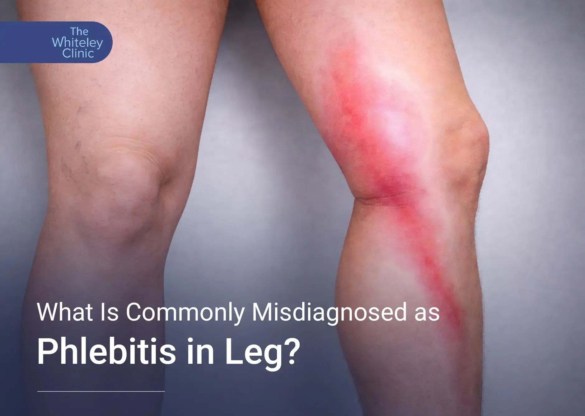
Recognising the Signs Before Complications Begin
Phlebitis in leg conditions can present in several distinct ways, ranging from mild local discomfort to visible inflammatory changes along the course of a vein. Although the condition follows recognised clinical patterns, not every patient experiences the same combination of symptoms. Understanding how phlebitis in leg develops, how it feels, and how it differs from other venous disorders is essential for accurate diagnosis and appropriate management.
Phlebitis in leg is an inflammatory condition affecting the veins, usually caused by a blood clot or injury to the vein wall. It can occur in both superficial and deeper veins, though it is most commonly seen in the superficial veins of the lower leg. The inflammation leads to pain, tenderness, and swelling along the affected vein, and in some cases, redness of the overlying skin. Recognising phlebitis in leg early is crucial because, while it is generally not life-threatening in superficial veins, untreated cases can sometimes progress or indicate underlying venous problems that require careful medical evaluation.
Read more: How Do Veins Work
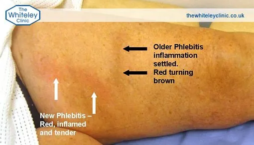
What Increases the Risk of Phlebitis?
An injection or cannula can sometimes irritate a vein. Pregnancy, smoking, obesity and hormonal treatments can also raise the risk of phlebitis, alongside conditions that make blood clot more easily. If episodes keep returning, the underlying cause may need investigating.
How Does Phlebitis Differ from DVT?
Superficial thrombophlebitis involves a clot in a vein just beneath the skin. DVT affects deeper veins. A clot in these veins may travel to the lungs and cause a pulmonary embolism. Because symptoms can overlap, looking at the leg is not enough to tell the two apart.
How Phlebitis in Leg Commonly Presents
As with many vascular conditions, phlebitis in the leg does not follow a single presentation. In clinical practice, symptoms may appear suddenly or develop gradually over one to two days. Some patients report an abrupt onset of pain, while others notice increasing tenderness that worsens over time.
Importantly, the absence of certain symptoms does not exclude phlebitis in the leg. Each case varies depending on the vein involved, its depth, and any underlying venous disease.
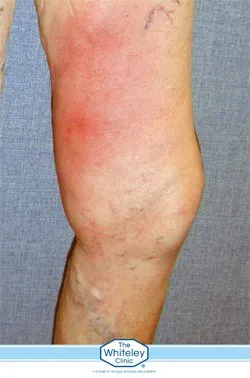
Early Symptoms: Localised Pain Along a Vein
The earliest symptom of phlebitis in the leg is usually discomfort, tenderness, or pain in one specific area of one leg. In rare cases, both legs may be affected. This pain is not diffuse. Instead, it follows the anatomical line of a vein.
Depending on the vein involved, pain may run:
- vertically along the inner thigh
- diagonally across the front of the upper thigh
- vertically along the inner calf
- vertically along the back of the calf
In patients with a previous history of varicose veins, phlebitis in the leg may appear as a series of tender lumps. These lumps often develop in the same locations as the earlier varicose veins.
Inflammation of the Vein and Overlying Skin Changes
Phlebitis in the leg occurs due to clot formation within a vein. As a result, the vein wall becomes inflamed. This inflammation does not remain confined to the vessel itself. Instead, it extends to the overlying skin.
Consequently, the skin above the affected vein usually appears red. This redness may not be visible if the vein lies deep beneath the skin. However, when the vein is superficial, tenderness is often marked. Pressure over the area typically increases pain, confirming local inflammation associated with phlebitis in the leg.
Palpable Findings: What Can Be Felt
When phlebitis in leg affects a superficial vein, physical examination often reveals a firm, tender structure beneath the skin. This structure feels like a hard, irregular “tube” and represents the inflamed vein containing a clot.
In contrast, when phlebitis in leg develops within varicose veins, the findings may differ slightly. Instead of a single cord-like structure, examination may reveal several rounded or diffuse lumps. Despite this difference, these areas remain firm to touch and painful when pressed.
Conditions Frequently Misdiagnosed as Phlebitis in the Leg
A significant clinical issue arises from the frequent misdiagnosis of phlebitis in the leg. Many inflammatory conditions of the lower leg are incorrectly labelled as phlebitis in the leg. This occurs when tenderness or redness prompts an assumption of venous thrombosis without proper assessment.
Such diagnostic errors matter. Incorrect labelling often leads to inappropriate treatment and delayed management of the true underlying condition.
Venous Skin Changes That Mimic Phlebitis
Several chronic venous disorders resemble phlebitis in leg in appearance but differ fundamentally in cause. These include:
- venous eczema (also known as varicose eczema)
- lipodermatosclerosis
- haemosiderin deposition
- venous leg ulcers
All these conditions arise from venous incompetence and venous reflux disease. Unlike phlebitis in leg, they do not involve clot formation within varicose veins or hidden varicose veins.
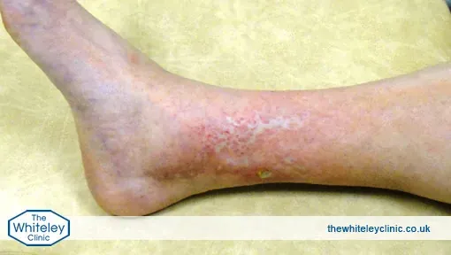
Understanding Venous Reflux and Valve Failure
In patients with venous incompetence, the valves within the leg veins fail to function correctly. When the leg relaxes, blood flows backwards down the veins instead of moving efficiently towards the heart.
This refluxing blood impacts the capillaries in the skin, particularly around the ankles. Over time, repeated pressure damages these tiny vessels. Unlike phlebitis in the leg, this process does not cause clots. Instead, it leads to cumulative micro-trauma within the skin and surrounding tissues.
Progressive Skin Damage Caused by Chronic Venous Disease
Initially, venous reflux causes little visible change. However, after years of repeated damage, inflammation develops around the capillaries. The skin begins to react in a predictable sequence.
First, irritation appears as venous eczema. Next, the skin becomes hardened and slightly swollen, a condition known as lipodermatosclerosis. Later, iron deposits accumulate, causing brown discolouration called haemosiderin deposition. Eventually, the skin breaks down, resulting in a venous leg ulcer.
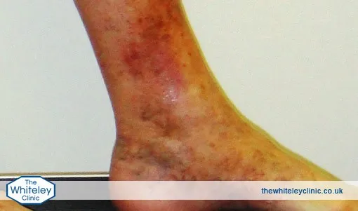
Atrophie Blanche: A Critical Warning Sign
Atrophie blanche represents an advanced stage of venous skin disease. In this condition, the skin around the ankle becomes thin, pale, and fragile. Breakdown into a venous ulcer often follows.
Although atrophie blanche may resemble phlebitis in leg to the untrained eye, it arises from venous incompetence and reflux disease, including hidden varicose veins. Crucially, it is not phlebitis in leg.
Why Correct Diagnosis Determines Correct Treatment
Management strategies differ significantly between phlebitis in leg and chronic venous disease. In venous eczema, lipodermatosclerosis, haemosiderin deposition, and venous ulcers, treatment must focus on stopping venous reflux. This requires proper investigation and management of venous incompetence.
By contrast, phlebitis in leg requires a different clinical approach because clot formation within the vein drives the inflammatory process. Confusing these conditions risks ineffective treatment and disease progression.
Seeking Advice About Your Symptoms
If you are concerned about phlebitis, contact The Whiteley Clinic team to ask about relevant services and appointment options.
New leg swelling, warmth or a hard, painful vein needs urgent medical assessment rather than waiting for a routine appointment. If leg pain or swelling comes with breathlessness or chest pain, seek emergency medical help immediately.
How Is Phlebitis Investigated?
A medical assessment usually starts with questions about your symptoms and medical history, followed by an examination. An ultrasound scan may be needed to check for a clot and whether deeper veins are involved. Treatment depends on what the assessment finds.
Treatment and Recovery
Once uncomplicated superficial phlebitis is confirmed, gentle walking, raising the leg when resting and a warm, moist cloth may help. Doctors can advise on pain relief. Do not take ibuprofen with prescribed blood thinners unless a doctor recommends it.
Compression stockings need professional assessment and fitting. Some cases require anticoagulants to reduce the risk of clot extension; DVT usually requires these medicines.
Mild superficial cases often improve within one to two weeks. Symptoms that worsen or fail to settle need reassessment. Regular movement supports circulation, although it cannot prevent every recurrence.