phlebitis
What Is Phlebitis and What’s the Best Treatment for It?
Phlebitis refers to inflammation of a vein, most often caused by a blood clot in a superficial vein. While it may seem like a minor issue initially, it often causes pain and discomfort. Furthermore, if left untreated, phlebitis can sometimes lead to more serious complications.
In this page, we explain what phlebitis is, explore its symptoms and causes, and discuss the best treatment options available to help you manage the condition effectively.
The term 'phlebitis' comes from two parts: 'phleb' meaning vein, and 'itis' meaning inflammation.
Although the term sounds simple, many people still misunderstand the condition. In reality, veins do not become inflamed without a reason. Most often, phlebitis develops when a blood clot forms in a vein close to the surface. When this occurs, the vein reacts by becoming inflamed to start the healing process.
Phlebitis in the Legs: What You Should Know
Phlebitis in the legs most commonly affects people with varicose or hidden varicose veins caused by venous reflux disease.
Normally, leg muscles contract during movement and push blood upwards through the veins. Then, when leg movement stops, the valves inside the veins quickly close to stop blood from flowing backwards. However, in cases of venous reflux, these valves fail. Consequently, blood flows back down the leg due to gravity, leading to a condition known as venous incompetence.
Over time, this backward flow leads to vein wall damage and dilation.
Once these dilated veins become visible under the skin, they are called varicose veins. However, when veins lie deeper and remain unseen, specialists refer to them as hidden varicose veins. Despite being out of sight, they behave just like surface varicose veins and carry similar risks.
What Causes Clot Formation?
In the 19th century, German physician Rudolf Virchow described three factors that contribute to clot formation, now known as Virchow’s Triad:
- Damage to the vein wall
- Changes in blood flow
- Changes in blood composition
In most cases, people with varicose or hidden varicose veins show two main contributing factors. First, the vein wall is damaged due to dilation. Second, blood flow is altered because of reflux. Consequently, clots may develop inside these abnormal veins.
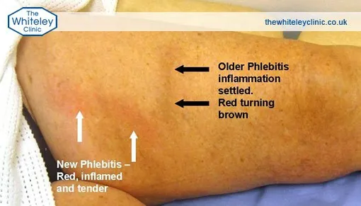
How Inflammation Develops
A blood clot forms inside a vein, a process known as thrombosis. This clot irritates the vein wall, leading to inflammation in the surrounding tissue. However, it is important to understand that this inflammation is not an infection; rather, it represents the body’s natural healing response. Consequently, increased blood flow brings white blood cells to the area, which gradually break down the clot over time.
Despite this, the clot may sometimes spread into deeper veins and lead to deep vein thrombosis (DVT). Even more concerning, a fragment of the clot may detach and travel to the lungs, resulting in a pulmonary embolism (PE). This condition poses a serious risk to life if not treated promptly by doctors.
A Misunderstood Condition: It’s Not an Infection
Many healthcare providers still confuse inflammation with infection. Since the affected area often feels hot, looks red, and is painful, they mistakenly prescribe antibiotics. However, bacteria do not cause phlebitis, so antibiotics are ineffective.
Instead, the inflammation is part of the body’s natural response to thrombosis. Unfortunately, misdiagnosis and incorrect treatment remain common.
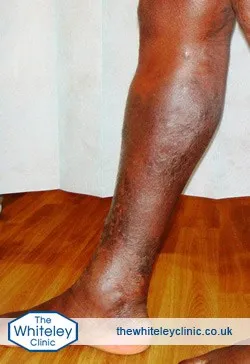
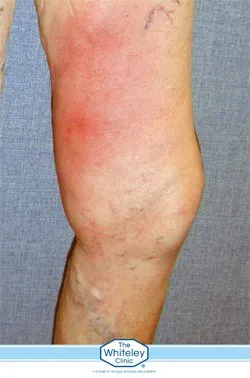
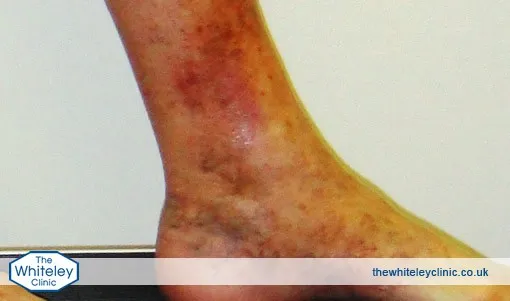
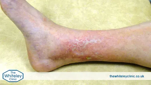
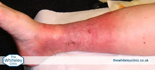
What Are the First Signs of Phlebitis to Watch For?
Common signs and symptoms include:
- A tender, red, and lumpy vein
- Warmth or pain in the affected area
- Localised swelling
- No signs of systemic infection (e.g., fever)
These symptoms usually occur just under the skin and should not be ignored.
What Happens If Phlebitis Is Left Untreated?
If phlebitis is not properly diagnosed and treated, the inflammation may spread from the superficial veins to the deep venous system. This progression increases the risk of developing deep vein thrombosis (DVT), a serious condition where clots form in deeper veins. In some cases, part of the clot can break off and travel to the lungs, causing a potentially life-threatening pulmonary embolism (PE). Early diagnosis through duplex ultrasound is essential to avoid these dangerous complications.
Studies in the UK and US have now confirmed that untreated phlebitis can lead to life-threatening complications.
A duplex ultrasound scan helps determine:
- Distance between the clot and the deep veins
- Whether treatment with anticoagulants is needed
- The underlying cause, such as reflux or varicose veins
Without a scan, it is impossible to know if the patient is at risk of DVT or PE. That’s why every case of phlebitis should be investigated with ultrasound.
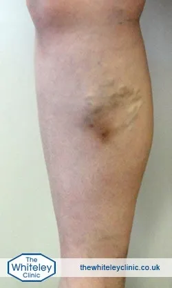
Can Phlebitis Go Away on Its Own?
In some cases, the inflammation and discomfort associated with phlebitis may gradually improve as the body naturally breaks down the clot. However, this does not mean the underlying problem has resolved. Without a duplex ultrasound scan, it is impossible to determine whether the clot is close to the deep venous system or whether there is an increased risk of deep vein thrombosis (DVT) or pulmonary embolism (PE). In addition, many cases of phlebitis are linked to underlying venous reflux or varicose veins, which may continue to cause problems if left untreated. For this reason, anyone with suspected phlebitis should seek specialist assessment rather than relying on symptoms to improve on their own.
Treatment Depends on the Scan
Without a duplex ultrasound scan, doctors cannot accurately decide the correct treatment for phlebitis.
This scan is essential to assess the exact location and extent of the clot, as appearances alone can be misleading. Only with this information can an effective treatment plan be put in place.
Once the ultrasound results are available, the approach becomes clear:
- If the clot is within 5 cm of a deep vein → Anticoagulation therapy (e.g., warfarin)
- Clot over 5 cm from deep veins → Aspirin and compression stockings recommended.
These treatments help reduce inflammation, limit clot progression, and lower the risk of complications.
To get an accurate diagnosis and proper treatment, Book a Free Discovery Call
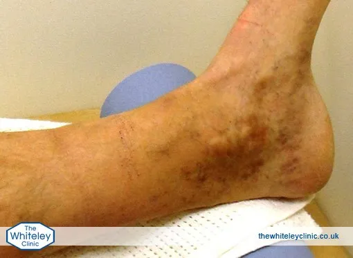
How Long Does Phlebitis Take to Heal?
With appropriate diagnosis and treatment, most cases of phlebitis begin to improve within a few days. Pain, tenderness and redness often start to settle during the first week, although the affected vein may remain firm or lumpy for several weeks while the body gradually breaks down the clot.
For many patients, symptoms improve significantly within 2–6 weeks. However, the exact recovery timeline depends on factors such as the size and location of the clot, whether it is close to the deep venous system, and the presence of underlying venous reflux or varicose veins.
At The Whiteley Clinic, treatment focuses not only on managing the inflammation and clot but also on identifying and treating the underlying vein problem. By addressing the root cause, recovery can be more effective and the risk of future episodes may be reduced. In some cases, complete resolution of vein hardness or residual tenderness can take several months, even though the main symptoms have already improved.
A duplex ultrasound scan is essential throughout the process, as it allows specialists to monitor the clot, assess healing and ensure there is no progression into the deep veins.
Key Facts About Phlebitis
- In phlebitis, the inflammation stems from a clot, not from an infection.
- Antibiotics are not effective.
- A duplex ultrasound plays a key role in diagnosis and assessing risk.
- Treatment plans depend on where the clot is and how far it has spread.
- Untreated cases may lead to serious problems, including DVT or PE.
- Hidden varicose veins often play a key role in causing phlebitis.
- Prevention requires accurate diagnosis and long-term vein care.
If you experience symptoms that may suggest phlebitis, it is important to seek medical attention from a specialist clinic. At The Whiteley Clinic, you can receive expert advice and the most effective treatments tailored to your condition. Timely diagnosis and appropriate care can prevent recurrence and protect your long-term health.