
Venous Eczema
Venous Eczema is a type of dermatitis linked to chronic venous disease. It usually affects the lower legs and may cause itchy, dry, scaly, red or inflamed skin, often around the ankle. It can develop alongside visible varicose veins, but visible veins are not always present. In some people, underlying venous reflux remains hidden beneath the skin. Accurate diagnosis therefore requires more than examining the skin. A specialist venous assessment and duplex ultrasound can identify the underlying venous problem and guide treatment. Managing the eczema itself can relieve symptoms, while treating significant venous reflux addresses an important underlying cause.
What Causes Venous Eczema?
The veins in the legs must return blood to the heart against gravity. During movement, the calf muscles help push blood upwards, while one-way valves help prevent it from flowing backwards.
When these valves become incompetent, blood can reflux down the vein. This increases venous pressure and can contribute to swelling, inflammation and changes in the skin. Over time, chronic venous disease may produce pigmentation, eczema and other forms of skin damage.
Venous Eczema therefore does not simply result from a problem on the surface of the skin. The skin changes can reflect an underlying problem with venous circulation.
The condition may occur with visible varicose veins. However, it can also occur when the abnormal veins cannot be seen from the outside.
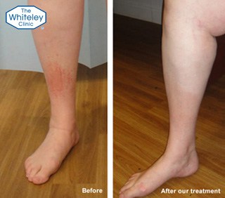
Can Hidden Varicose Veins Cause Visible Eczema?
Yes. Venous reflux can exist without obvious bulging veins on the surface of the leg.
Visible varicose veins represent one clinical sign of chronic venous disease. However, reflux can also affect veins deeper within the leg or involve incompetent perforator veins that are not obvious during a visual examination.
This creates an important diagnostic point. A person may have ankle eczema, swelling or other venous skin changes without having visibly prominent varicose veins.
Duplex ultrasound can identify abnormal venous flow and show which veins contain reflux. NICE recommends duplex ultrasound to confirm varicose veins and assess truncal reflux when planning treatment.
At The Whiteley Clinic, the Whiteley Protocol® uses a detailed venous duplex assessment to map the veins contributing to the problem. The clinic states that this assessment examines the veins of both legs and can include specialist pelvic vein assessment when indicated.
What Does Venous Eczema Look Like?
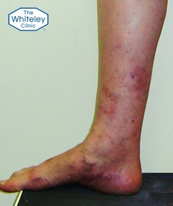
Venous Eczema can vary in appearance. Common features include:
- Red or pink areas of inflamed skin
- Itching or irritation
- Dry, scaly or rough skin
- Crusting or weeping in more active eczema
- Swelling around the ankle or lower leg
- Brown or orange-brown pigmentation caused by haemosiderin
- Skin thickening in longer-standing venous disease
The condition often affects the lower leg, particularly around the ankle. It may appear as a defined patch or spread across a larger area of the leg.
The appearance alone cannot confirm the diagnosis. Several skin conditions can cause similar changes on the legs, including contact dermatitis, psoriasis, asteatotic eczema and other inflammatory disorders.
Is Venous Eczema a CEAP C4 Condition?
Yes. Within the clinical component of the CEAP classification, eczema caused by chronic venous disease falls under C4a.
CEAP classifies chronic venous disease according to clinical, aetiological, anatomical and pathophysiological features. The clinical classes progress from C0, with no visible signs, to C6, which represents an active venous leg ulcer.
The relevant stages include:
- C2 – varicose veins
- C3 – oedema
- C4a – pigmentation or eczema
- C4b – lipodermatosclerosis or atrophie blanche
- C4c – corona phlebectatica
- C5 – healed venous ulcer
- C6 – active venous ulcer
Therefore, Venous Eczema represents a skin manifestation of more advanced chronic venous disease rather than a purely cosmetic skin problem.
What Does Eczema in Different Areas of the Leg Mean?
The location of eczema can provide useful clues, but it cannot establish the cause on its own.
Venous Eczema most commonly affects the lower leg, particularly around the ankle. If eczema appears mainly around the knee, the pattern is less typical of venous disease. Other diagnoses may need consideration, depending on the appearance, distribution and other symptoms.
Eczema elsewhere on the body can also change the diagnostic picture. For example, similar patches on the arms, scalp or other areas may suggest a broader dermatological condition rather than a problem caused solely by venous disease.
Other causes of leg dermatitis include psoriasis, contact dermatitis, atopic dermatitis, nummular dermatitis and asteatotic eczema. A clinical assessment can help distinguish these conditions from venous skin disease.
How Is Venous Eczema Diagnosed?
A diagnosis should consider both the skin and the underlying venous circulation.
A clinical examination can identify signs such as varicose veins, oedema, pigmentation and skin changes. However, visual examination cannot reliably exclude venous reflux when no veins are visible.
A venous duplex ultrasound provides a more detailed assessment. It can show the direction of blood flow and identify incompetent veins contributing to venous hypertension.
The quality and scope of the scan also matter. The Whiteley Protocol® places detailed duplex ultrasound at the beginning of the diagnostic process. According to The Whiteley Clinic, the assessment maps the veins of both legs and can extend to the pelvic veins when the clinical picture indicates a pelvic source.
This approach can help identify visible and hidden sources of venous reflux before treatment is selected.
Are Steroid Creams a Permanent Treatment?
No. Topical corticosteroids can help control the inflammatory skin component of Venous Eczema, particularly during a flare. Emollients can also help restore the skin barrier and reduce dryness.
However, these treatments do not correct an incompetent venous valve or stop venous reflux.
For this reason, topical treatment and treatment of the underlying venous disease serve different purposes. Skin treatment can control symptoms, while treatment of significant venous reflux aims to address the circulatory cause.
Topical corticosteroids should therefore be used appropriately and for the duration recommended by a clinician. Prolonged or inappropriate use can cause adverse effects, including skin thinning.
Are Compression Stockings a Permanent Cure?
Compression has an established role in managing venous disease. It can reduce oedema and improve venous return, which may also reduce venous skin symptoms.
However, compression does not remove the underlying source of venous reflux. Its effect depends on continued use.
This distinction is particularly important when the aim is to treat the cause rather than simply control symptoms. NICE recommends compression hosiery for varicose veins when interventional treatment is unsuitable, rather than as a replacement for appropriate intervention when treatment is indicated.
Therefore, compression can form part of management, but it should not automatically be regarded as a permanent solution for the underlying venous problem.
How Is Venous Eczema Treated?
Treatment depends on the venous pattern identified during investigation.
The first step is to identify the veins responsible for reflux. Once the anatomy and direction of abnormal blood flow are established, treatment can target the affected veins.
Modern venous treatment includes minimally invasive techniques such as endovenous laser ablation, ultrasound-guided foam sclerotherapy and treatment of incompetent perforator veins. NICE recommends endothermal ablation for suitable patients with confirmed varicose veins and truncal reflux, with ultrasound-guided foam sclerotherapy or surgery considered when endothermal treatment is unsuitable.
At The Whiteley Clinic, treatment follows the individual venous pattern identified through The Whiteley Protocol®. The clinic currently describes a staged approach. The first stage aims to stop significant venous reflux. Depending on the veins involved, this may include endovenous laser ablation, HIFU, TRLOP for incompetent perforators, pelvic vein embolisation or ambulatory phlebectomy.
The second stage can involve ultrasound-guided foam sclerotherapy to treat remaining refluxing or smaller veins. The clinic also uses the VARIXIO® system for foam preparation. A further cosmetic stage may be considered for residual surface veins when appropriate.
The exact combination should therefore follow the ultrasound findings rather than the appearance of the skin alone.
Why Accurate Venous Mapping Matters
Venous disease can involve more than one type of vein. Reflux may affect truncal veins, perforators, tributaries or, in selected cases, pelvic veins.
This means that treating one visible varicose vein may not address every source of abnormal venous flow.
The Whiteley Protocol® uses a structured approach to investigate and treat venous disease. It starts with detailed duplex ultrasound to map the venous system before the clinician selects a treatment. The treatment then targets the veins responsible for reflux.
The clinic has reported cases where Venous Eczema occurred alongside truncal reflux and incompetent perforators. In one published case, clinicians used endovenous laser ablation, TRLOP and ambulatory phlebectomy after completing a detailed duplex assessment.
Can Treating the Veins Improve Venous Eczema?
Treating the underlying venous problem can reduce the venous pressure and inflammation contributing to skin changes.
The outcome depends on the severity and duration of the skin disease, the extent of venous damage and the presence of other conditions. Earlier assessment may help prevent progression to more advanced skin changes.
Venous disease can progress from oedema and eczema to more established skin damage and, in some patients, venous ulceration. The CEAP classification reflects this progression from C4 skin changes to C5 healed ulcer and C6 active ulcer.
Persistent eczema around the ankle or lower leg may have a venous cause, rather than a skin condition alone. Doctors should investigate venous disease when eczema occurs alongside swelling, aching, pigmentation or varicose veins.
When Should You Seek Help for Venous Eczema?
Persistent or recurrent eczema on the lower legs deserves assessment when it occurs with ankle swelling, varicose veins, leg heaviness, aching, itching or brown skin pigmentation.
The absence of visible varicose veins does not exclude venous disease. Similarly, improvement with steroid cream or compression does not prove that the underlying cause has been resolved.
A specialist assessment can establish whether venous reflux contributes to the skin changes and whether further treatment is appropriate. When significant reflux is present, addressing the underlying venous disease can form an important part of long-term management.