
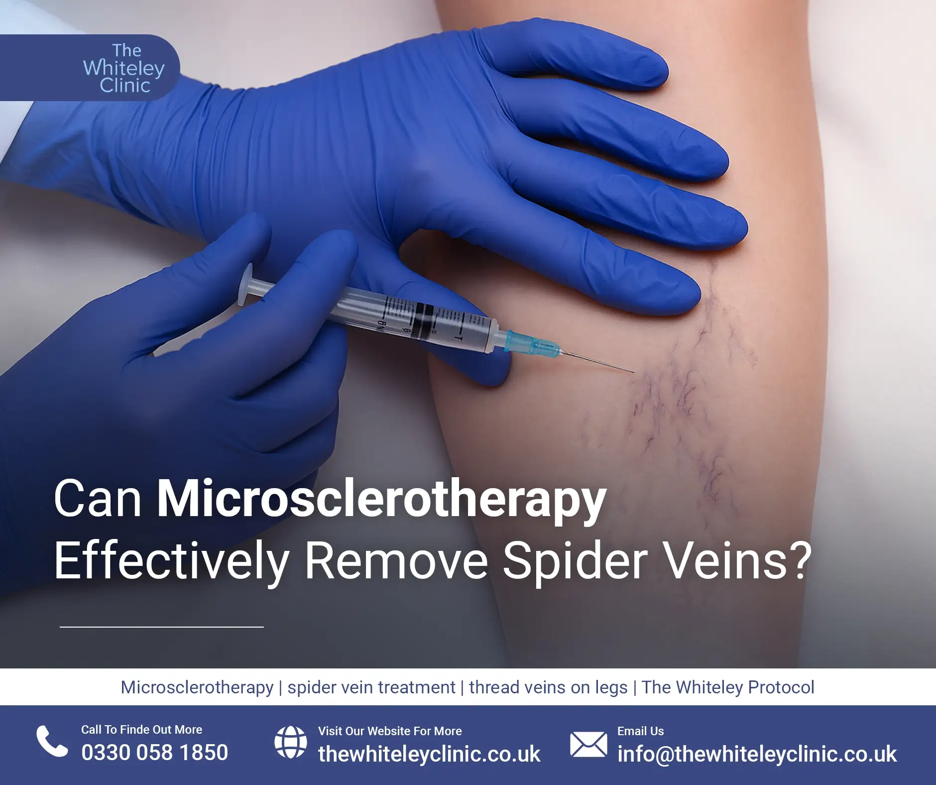
Can Microsclerotherapy Effectively Remove Spider Veins?
July 18, 2026
Up until the invention of venous Duplex Ultrasound scanning there were several different methods for the diagnosis of varicose veins. As you can see from the NICE guidelines, Duplex Ultrasound scanning has rightly become the gold standard diagnosis technique for the investigation of varicose veins.
However, prior to the introduction of Duplex Ultrasound, there existed various different tests for varicose veins. Many of these tests are now considered obsolete; but some of them can still add value to specific diagnosis and treatments in certain cases.
Please be aware that the clinic does not store Ultrasound images for routine venous leg scans.
We routinely take images for specific Ultrasound scans, which require classic reproducible views and measurement data to be recorded. These include pelvic congestion/transvaginal scans, male pelvic/testicular scans, arterial scans, deep vein thrombosis scans. Only this type of image can be requested after completing a Subject Access Request (SARs) form, which you can request at reception.
Click the titles below or use the side menu for more information about each technique
This technique forms the foundation of all our testing under The Whiteley Protocol®. Every patient will be examined using this technique as the starting point for any investigation.
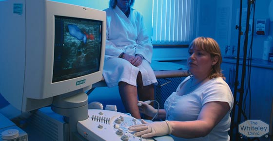
The word ‘duplex’ refers to the two different elements involved in this kind of scan, although in reality there are technically three stages involved. In fact, in some parts of the world, it is referred to as ‘triplex scanning’, ‘colour-coded Duplex scanning’ or ‘colour flow Duplex scanning’.
The initial process involved in venous Duplex Ultrasound scanning is a ‘greyscale/B-mode ultrasound’ scan… the standard sort of ultrasound scan that we’re all accustomed to seeing, when doctors apply gel to a pregnant woman’s bump and roll a scanner to see the baby in the womb. Ultrasound scanners do not produce any radiation (unlike x-rays) and they don’t involve needles or injections.
The second part of the process is the ‘Doppler ultrasound’ scan, beamed into one specific area of the body and capable of being directed inside the vein. By combining the ultrasound picture generated with the specific Doppler waveform, Doppler ultrasound can be used to measure blood flow in the vein.
Finally, the third part of the scanning process is the ‘colour flow’/‘colour-coded’ element. Computers these days are so fast that any movement on the black and white ultrasound picture can be picked up and ‘coded’ as either blue or red depending on the direction of flow, enabling blood flow to be seen in a vein in real time. In addition, we can see when blood flows up the vein normally and when it flows down the vein abnormally if the valves are not working.
It is this ability of the Duplex Ultrasound scan to actually see the flow in real life, that makes it such an incredibly powerful technique in the investigation of every individual patient’s varicose vein problems and in the determination of which treatment – or combination of treatments – is best for that patient.
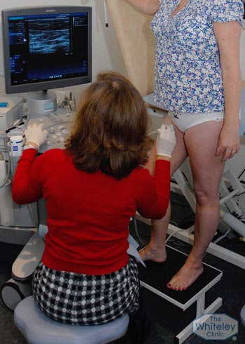
It’s worth remembering that a venous Duplex Ultrasound scan is only as good as the person performing it. To ensure optimum results, the scan should be performed by a specialist vascular scientist who scans veins every day, as is the case at The Whiteley Clinic. It is a very specialised and detailed process, which should take between 30 to 45 minutes per scan to receive an accurate diagnosis.
The kind of scans undertaken by many other clinics – using small machines, taking just 5 to 10 minutes, and often performed in a rush by the doctors themselves or by less qualified personnel – will often miss important underlying causes of varicose veins.
At The Whiteley Clinic, a full ‘Gold Standard’ Duplex Ultrasound is our ‘Minimum Standard’ for obtaining a full and comprehensive diagnosis. However, there are some things that even the most professionally conducted Duplex Ultrasound scan is not able to show, and for that reason, we may recommend additional diagnosis methods such as Intravascular ultrasound or Plethysmography.
Plethysmography is a technique that measures the changes in volume of a limb. There are three principal means of applying the technique: photo plethysmography, air plethysmography and strain gauge plethysmography.
In photo plethysmography (PPG), an infrared light probe shines infrared light into the capillaries and measures the reflection. As the venous pressure rises and the veins dilate, the speed of dilation can be measured. In air plethysmography (APG) and strain gauge plethysmography, the volume of the leg (usually the calf) is measured. The leg is usually elevated and dropped to see how the movement of blood in the veins changes the volume of the limb.
From these results, we are able to observe the function of veins as a whole rather than just which way the blood is flowing, as observed with venous Duplex Ultrasound scanning. Thus, the information provided by plethysmography adds to the information that can be gleaned from venous Duplex Ultrasound scanning and we quite often perform both techniques together to obtain a full assessment of the venous disease in a leg.
This is a fantastic new technology, available to The Whiteley Clinic, in which a small ultrasound probe can be passed into a vein on the end of the catheter. It is very useful for looking at veins that are narrowed or being compressed and so, as with CT and MRI, it may be useful in post thrombotic syndrome (PTS). However, in the investigation of normal varicose veins, IVUS is not required.
Research from The Whiteley Clinic has shown that varicose veins for 1 in every 7 women actually come from the pelvis and into the legs. This increases to 1 in every 5 women who have had children.
If a duplex ultrasound scan indicates that the varicose veins are coming from the pelvis, it is important to understand which of the veins in the pelvis is causing the problem. Due to their location, these veins are more difficult to see and so a transvaginal duplex ultrasound scan needs to be undertaken.
This technique, which was developed at The Whiteley Clinic, is relatively new and not widely available elsewhere.
As missing pelvic varicose veins is a major cause of recurrent varicose veins, it is imperative that they are correctly identified and treated.
The technique of venography involves the injection of a contrast solution into a vein followed by a series of x-rays to watch the process of venous reflux as the contrast solution flows up inside the vein. It has been used in conventional medical practice as a diagnostic tool. However, at The Whiteley Clinic, our research has proven categorically that Duplex Ultrasound is by far the more accurate diagnostic technique. For that reason, we reserve the use of venography as part of the treatment to make sure any coil embolisation or foam embolisation is positioned exactly as required. We may occasionally also use this technique in the specific diagnosis of male pelvic vein problems, and to guide treatment catheters for female pelvic vein embolization following a diagnosis using the far superior transvaginal Duplex Ultrasound scanning.
CT and MRI technology provide excellent scans for a great many areas of medicine.
And whilst they are virtually never needed as a part of day-to-day varicose vein surgery under The Whiteley Protocol®, they can nonetheless be useful in deep vein thrombosis or in people with severe venous disease such as in post thrombotic syndrome (PTS).
Historically, before Doppler ultrasound was available and before we understood the underlying mechanisms of varicose vein development, the Trendelenburg or ‘tourniquet’ test was used to check the source of varicose veins. Now relegated to the dustbin of history, this technique is considered to be completely inaccurate compared to Duplex Ultrasound.
Another historical curiosity, small hand held ‘pencil Dopplers’ were common equipment in every hospital and venous practice in the 1980s. Small enough to fit in a pocket and never intended to connect to a TV monitor, these devices would be held at an angle next to the skin and would ‘beep’ if a blood flow was detected. Not surprisingly, by the late 1990s, hand-held Dopplers had come to be regarded as inaccurate at best, at worst misleading and entirely inadequate for the investigation of varicose veins and venous reflux. You might occasionally see one being used to measure arterial pressure… but not at The Whiteley Clinic!