
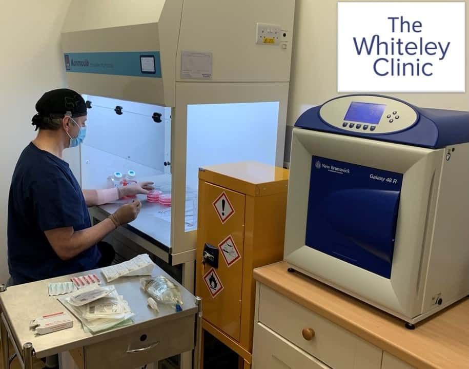
Varicose vein research laboratory
February 19, 2026
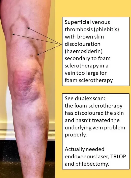
This lady attended The Whiteley Clinic in July 2016 asking for a second opinion. She had a large discoloured area of her left calf. She thought this looked like a bruise (see picture). Over the last eight years, she has had several treatments for spider veins of the legs in this area. The first was performed by a doctor who used a needle that generated heat by using an electric current. This did not work and indeed the area seemed to get worse.
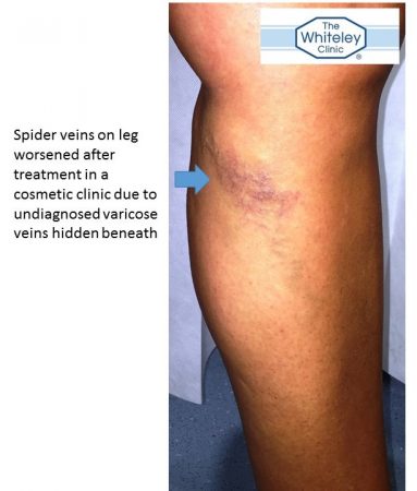
Leg spider veins worse after treatment due to hidden varicose veins
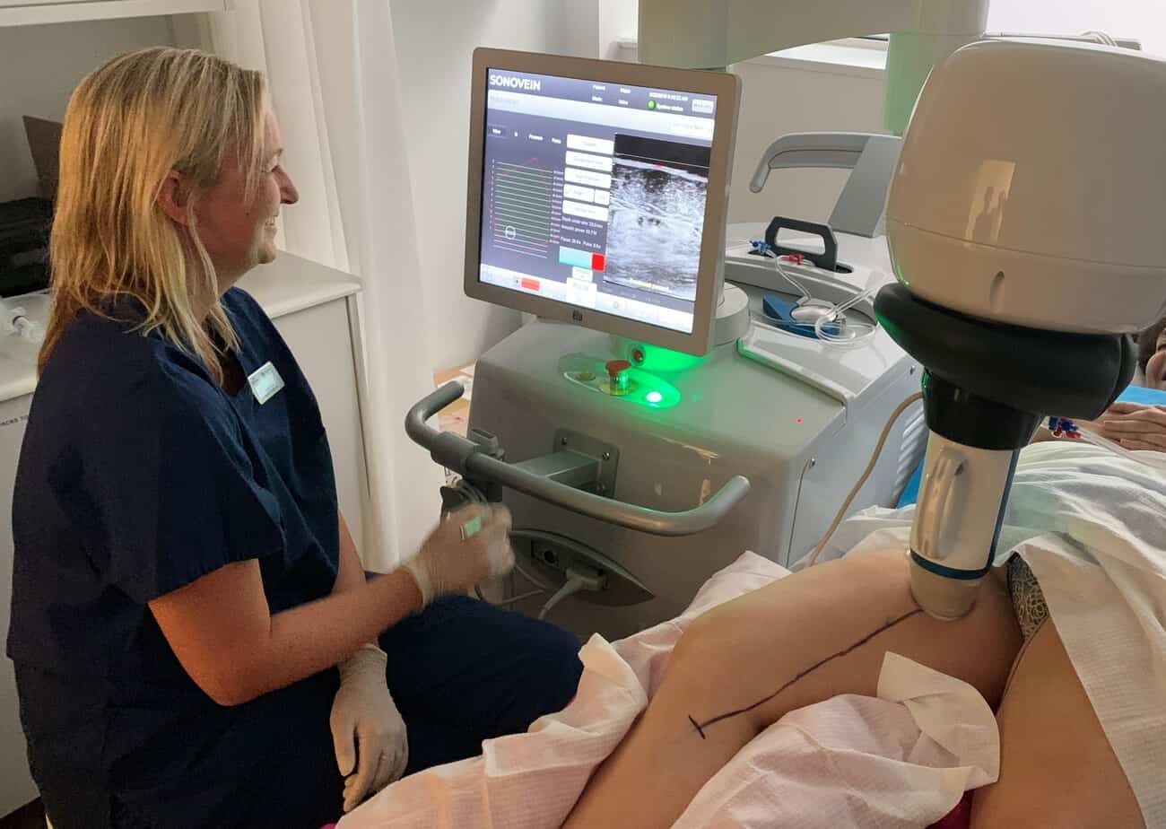
Subsequently, she went to a cosmetic clinic where these veins were injected with sclerotherapy. Once again, not only did the spider veins remain, but they worsen significantly. She came to The Whiteley Clinic having read The Whiteley Clinic website and realise that she had never been offered a venous duplex ultrasound scan before any of her treatments. Quite correctly, she realised that no one had checked for underlying "hidden varicose veins" before treating her. At The Whiteley Clinic she underwent a venous duplex ultrasound scan performed by a The Whiteley Clinic trained vascular technologist. This scan is much more detailed than many scans performed by doctors or nurses themselves. It takes approximately 20 minutes per leg to get a good understanding of the individual pattern of disease. 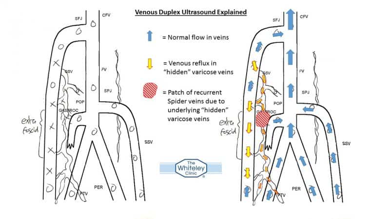 Venous Duplex Ultrasound showing varicose veins underlying - spider veins thread veins - The Whiteley Clinic
The venous duplex ultrasound scan is shown here (see picture of scan). On the left is the original scan and on the right coloured arrows have been added to explain where the hidden varicose veins are. All of the blue arrows show normal flow, but the red and yellow arrows show the hidden varicose veins. In these veins, which are not visible on the surface, the valves have failed. On standing, blood falls the wrong way down these veins. In this lady's case, at one point these come close to the skin. Because of this blood flowing the wrong way, the veins start to bulge and where they come near the skin, a patch of spider veins has emerged. Because she had not had this scan before, it is not surprising that her spider veins had worsened considerably after treatment with the needle heating the tissue with electric current and the sclerotherapy. Both of these techniques can destroy small veins, but if there is a large amount of blood refluxing under them from hidden varicose veins, then it can actually worsen the situation as in this case. The optimal management for leg spider veins as illustrated by this lady is: 1] get a proper venous duplex ultrasound scan performed by an expert clinic to check for underlying hidden varicose veins 2] have any underlying hidden varicose veins treated before treating the spider veins 3] if the spider veins remain after treating the underlying varicose veins, then treat them. In many cases, treating the hidden varicose veins will get rid of the spider veins at the same time.
There is a reasonable amount of research that has now been performed into this question. Approximately 89% of women with spider veins of the legs have significant underlying hidden varicose veins. About 40% of women with leg spider veins have severe underlying hidden varicose veins and 15% have incompetent perforating veins. With the amount of evidence now available, it seems incredible that patients allow doctors, nurses or therapists to treat their leg spider veins without getting a specialist venous duplex ultrasound performed first. It is even more incredible that professional clinics do not offer this as part of the service.