

Klippel-Trenaunay Syndrome (KTS)
March 26, 2026
Judy Holdstock of The Whiteley Clinic won the first prize at the American College of Phlebology annual meeting in Austin, Texas this weekend.
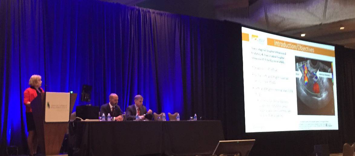 Judy Holdstock giving her research presentation on the extended pelvic veins scan which went on to win First Prize at the American College of Phlebology 2017
Her prize-winning research showed how a pelvic veins scan, performed with duplex ultrasound and the Holdstock protocol shows more than other current tests. Duplex ultrasound scanning is done lying flat, or just across the abdomen, or MRI, CT or venograms, have all overestimated a pelvic vein problem called "Nutcracker" phenomena. This has meant that in the past, many patients (mainly women) with pelvic congestion syndrome or other pelvic varicose veins problems, have been refused curative treatment or potentially offered surgery or stenting rather that curative pelvic vein embolisation with coils.
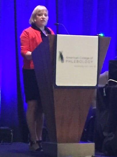
Judy Holdstock presenting at The American College of Phlebology 2017 - Winner of First Prize of meeting
Just like veins of the legs can cause leg varicose veins, pelvic veins can cause pelvic varicose veins. These pelvic varicose veins can cause pelvic congestion syndrome and/or varicose veins of the vulva, vagina and buttocks in females. In males, the same veins can cause a varicocele. This is a varicose vein around the testicle. In both males and females, pelvic varicose veins can also cause leg varicose veins. Failure to diagnose and treat the pelvic varicose veins can mean failure to treat pelvic congestion syndrome or leg varicose veins that keep coming back again despite varicose vein treatments to leg veins.
The optimal way to investigate the pelvic veins is still controversial. Some doctors use MRI, CT scanning or venogram. However, research suggests that these techniques are inferior to transvaginal duplex ultrasound scanning when performed using the Holdstock technique. Judy Holdstock of The Whiteley Clinic developed the Holdstock technique for scanning pelvic veins. Previous research has suggested this is the gold standard test for pelvic vein reflux. This appears to be the cause of pelvic congestion sent as well as some of the varicose veins of the vulva, buttocks and legs.
Many doctors believe that pelvic varicose veins are due to narrowings or blockages of veins in the pelvis. These are often called “Nutcracker phenomenon" when the left renal (kidney) vein is narrowed. If there is pain in the left flank and microscopic blood in urine, then the "Nutcracker phenomenon" is called "Nutcracker syndrome". Many experts in the USA and Europe diagnose "Nutcracker phenomenon" because they see a narrowing of the left renal vein on ultrasound, MRI, CT scan or venography. However, Judy's research has shown that by using her protocol and then checking patients after embolisation of the left ovarian vein, the Nutcracker phenomenon disappears. She has shown that the apparent narrowing using normal tests is not the cause of the problem, but an effect of having left ovarian varicose veins. When these are treated by pelvic vein embolisation, this worrying appearance disappears.

Judy Holdstock presented with First Prize at the American College of Phlebology 2017 - at after conference party
Judy Holdstock and her team of vascular scientist at The Whiteley Clinic have developed the extended pelvic vein scan. The patient is scanned in several positions, on their backs, on their sides and at an angle of between 30 and 45° to allow the blood to reflux back down any incompetent veins with gravity. The pelvic veins are examined low in the pelvis using a transvaginal scan with the patient at this angle, and the veins in the upper abdomen and flank are examined from externally. Judy's research has shown that by using this extended pelvic vein scan, she and her team are able to diagnose pelvic vein problems that have previously been misdiagnosed by all other tests. By using Judy Holdstock's extended pelvic veins scan, patients at The Whiteley Clinic are ensured of getting the most accurate diagnostic scan of their pelvic veins available at the current time and are therefore more likely to get the right treatment for their problem and the highest chance of a cure.
Judy Holdstock won this coveted international prize for her and her team showing how the Holdstock extended pelvic veins scan can be used to accurately diagnose pelvic vein problems, and show that other doctors have been over-diagnosing the "Nutcracker phenomenon". We congratulate Judy on winning this prize for her years of dedicated work in this area that translates to patients getting the very best diagnosis and treatment available.
Pelvic congestion syndrome Pelvic veins scan Pelvic vein embolisation