
Haemosiderin (Brown Stains)
Brown stains on the legs can be more than a cosmetic concern. Known medically as Haemosiderin staining, these brown areas often develop around the ankles and lower legs when chronic venous disease affects the skin. Brown stains may therefore indicate an underlying problem with venous blood flow. This article explains what Haemosiderin is, why Brown Stains develop, how they relate to varicose veins and chronic venous insufficiency, and how the underlying vein problem and skin changes can be treated.
What Are Brown Stains on the Legs?
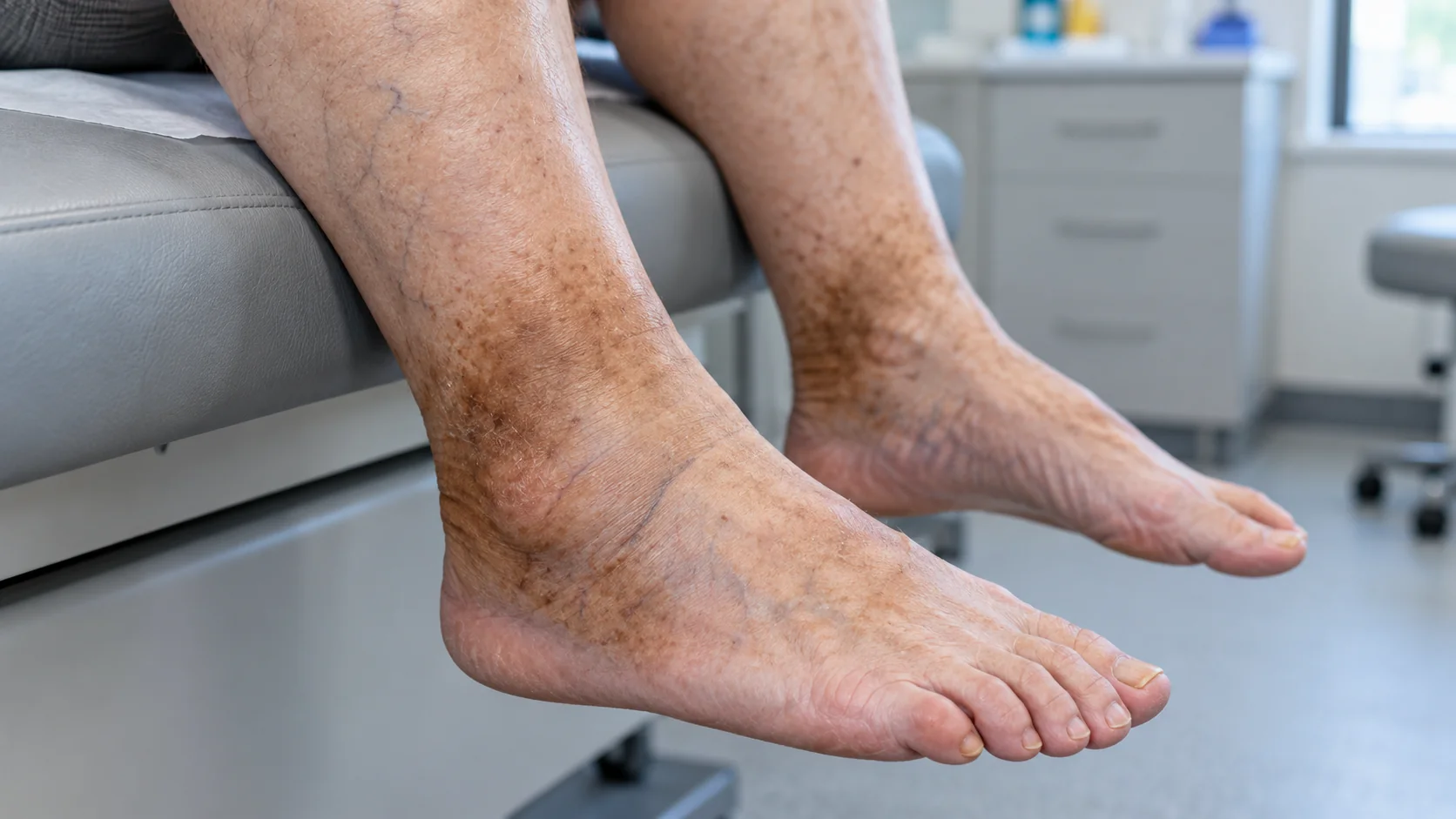
Brown stains are areas of darker skin that commonly appear around the ankle or lower calf. The colour develops when red blood cells leave small blood vessels and break down within surrounding tissue.
As these cells break down, iron is released. The body stores some of this iron within a protein complex called Haemosiderin. Over time, this can create a persistent brown or reddish-brown appearance in the skin.
The European Society for Vascular Surgery recognises brown pigmentation as a sign of more advanced chronic venous disease. Other changes can include swelling, eczema, hardened skin and venous ulceration.
Why Does Haemosiderin Develop?
The main cause of Haemosiderin staining is chronic venous hypertension, which often results from venous reflux.
Healthy leg veins contain valves that help blood return to the heart. When these valves stop working properly, blood flows backwards and increases pressure in the lower leg, especially when standing.
Over time, increased venous pressure can damage small blood vessels around the ankle. Red blood cells may then escape into the surrounding tissue. When these cells break down, they release iron that accumulates in the skin and contributes to Brown Stains.
Common Causes of Haemosiderin Staining
Several conditions can contribute to Haemosiderin deposition:
- Varicose veins: Damaged venous valves can cause blood to flow backwards and increase pressure in the leg.
- Hidden varicose veins: Reflux can occur in veins that a simple visual examination cannot detect.
- Previous venous leg ulcers: Healed ulcers may leave persistent skin pigmentation.
- Chronic inflammation: Long-term inflammation can contribute to tissue damage and iron deposition.
- Previous sclerotherapy: Some patients develop skin pigmentation following sclerotherapy.
- Leg infections: Certain infections can cause inflammation and changes in skin colour.
Not every brown mark has the same cause. A specialist should assess the skin changes and investigate any underlying venous problem before recommending treatment.
Can Varicose Veins Cause Brown Stains?
Yes, Brown Stains can occur as a consequence of chronic venous disease associated with varicose veins.
NICE considers pigmentation caused by chronic venous insufficiency a reason for referral to a vascular service. The guideline also recommends duplex ultrasound to confirm venous disease, identify reflux and help plan treatment.
This matters because treating the visible veins alone may not address every source of abnormal blood flow. A detailed assessment can identify reflux in the main superficial veins, tributaries or other veins contributing to the problem.
At The Whiteley Clinic, The Whiteley Protocol includes this assessment. A specialist performs venous duplex ultrasound to identify the underlying venous problem before planning treatment.
What Does Haemosiderin Mean in the CEAP Classification?
The CEAP classification describes the clinical severity and characteristics of chronic venous disease.
The later stages include a healed venous leg ulcer at C5 and an active venous leg ulcer at C6. NICE defines C4 as skin changes such as pigmentation, eczema, lipodermatosclerosis or atrophie blanche.
Therefore, Haemosiderin staining should not automatically be viewed as an isolated cosmetic problem. When venous reflux causes the pigmentation, it represents a visible sign of chronic venous disease.
Can Brown Stains Lead to a Leg Ulcer?
Brown pigmentation does not mean that an ulcer will definitely develop. However, it can occur alongside other signs of advanced venous disease.
Chronic venous hypertension can progressively damage the skin and underlying tissues. NHS guidance lists skin discolouration, swelling, hardened skin and varicose eczema among features associated with venous leg disease. Venous leg ulcers commonly develop around the lower leg and ankle when chronic venous problems damage the skin.
For this reason, new Brown Stains should not simply be covered with cosmetic products without investigating the possible venous cause.
How Are Brown Stains Diagnosed?
A clinical examination provides the first step. A specialist may assess the skin, visible veins, swelling and other signs of chronic venous disease.
A venous duplex ultrasound can then assess blood flow and identify venous reflux. NICE specifically recommends duplex ultrasound for people with suspected primary or recurrent varicose veins.
This investigation is particularly important when the skin changes appear without obvious varicose veins. In such cases, underlying reflux may not be visible from the surface.
The diagnosis should also consider other causes of skin pigmentation. Not every brown patch on the leg represents Haemosiderin.
Can Haemosiderin Be Treated?
Yes. However, treatment usually requires two separate considerations.
First, a vascular specialist should assess any underlying venous reflux and determine whether treatment is necessary. NICE recommends endothermal ablation for suitable patients with confirmed varicose veins and truncal reflux. If endothermal ablation is unsuitable, clinicians can consider ultrasound-guided foam sclerotherapy. When neither option suits the patient, clinicians may recommend surgery.
The Whiteley Clinic uses treatments such as endovenous laser ablation, radiofrequency ablation, TRLOP, phlebectomy and foam sclerotherapy according to the individual pattern of venous disease. The appropriate combination depends on the veins involved and the findings of the venous assessment.
Treating the reflux addresses the underlying cause. However, the existing Haemosiderin deposits may not disappear immediately after successful vein treatment.

How Are Brown Stains Treated?
Once the underlying venous problem has been assessed and treated where necessary, the remaining Brown Stains may require separate treatment.
The Whiteley Clinic uses a combination approach involving specific laser treatment and a topical skin-care regime for suitable patients. The aim is to target the Haemosiderin deposits responsible for the visible pigmentation.
The number of treatment sessions varies between patients. It can depend on the size of the affected area and the depth or darkness of the pigmentation.
This means that treatment should not focus on skin colour alone. The underlying venous circulation must first be assessed, particularly when the staining occurs around the ankle or lower leg.
What Can You Do If You Notice Brown Stains?
If Brown Stains develop around the ankle or lower leg, particularly alongside varicose veins, swelling, aching or heaviness, a medical assessment is appropriate.
NHS guidance also recommends medical assessment when chronic venous skin problems develop. Varicose eczema, for example, can cause colour changes, swelling, itching and skin thickening, and untreated skin damage can contribute to ulcer formation.
Keeping physically active and avoiding prolonged periods of standing or sitting may help venous symptoms. Leg elevation can also help reduce swelling. However, these measures do not replace assessment of significant venous reflux.
If an open sore develops on the lower leg and does not heal within two weeks, it may represent a venous leg ulcer and requires specialist assessment.
Treat the Veins, Not Just the Skin
Haemosiderin staining can be the visible result of a much deeper venous problem. For this reason, treating the colour alone may not provide a complete solution.
A proper assessment should establish whether venous reflux is present and determine which veins are responsible. Where appropriate, treatment can then address the underlying venous disease before the remaining Brown Stains are treated.
At The Whiteley Clinic, this approach forms part of The Whiteley Protocol. It combines detailed venous assessment with an individual treatment plan and, where appropriate, specific treatment for residual skin pigmentation.
The goal is therefore not simply to make the skin look clearer. It is to identify and manage the venous problem responsible for the skin changes, while also addressing the remaining Haemosiderin staining where suitable.