Hidden Varicose Veins
The term “hidden varicose veins” appears to have been first used in 2011. Prof Mark Whiteley use the term to explain venous reflux in legs without any
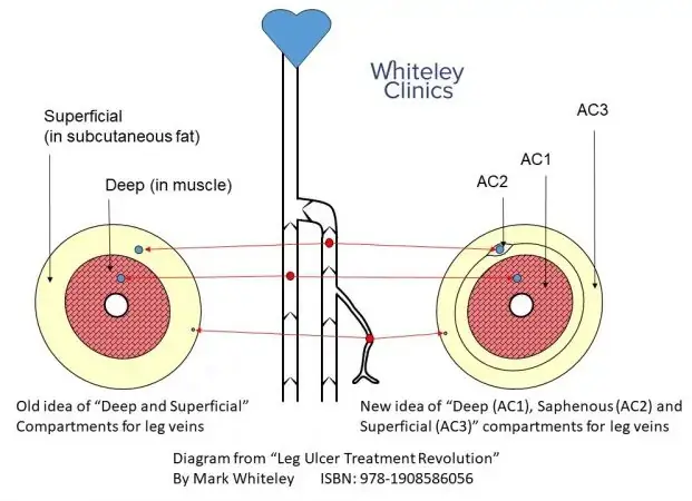
Superficial and deep veins of the legs
Before we can fully understand varicose veins and hidden varicose veins, we have to understand a little about the anatomy of the leg veins.
Most doctors and nurses still think of veins being either deep or superficial. Deep veins are in the muscle. Superficial veins are in the subcutaneous fat under the skin.
Superficial, saphenous and deep veins of the legs
Unfortunately, this is wrong. An Italian doctor called Paolo Zamboni has popularised a much more accurate understanding of the leg veins. The deep veins are in the muscle. These are the network one veins (N1). The muscle compartment is the anatomical compartment 1 (AC1).
The main superficial veins in the legs are the saphenous veins. There is the great saphenous vein (GSV) and small saphenous vein (SSV). The old names are the long saphenous vein and the short saphenous vein. However, these names were changed in Europe in 2001-2 and America in 2004-5. Healthcare professionals using these old terms are well over a decade out of date.
In addition, a third main vein is called the anterior accessory saphenous vein (AASV). These three saphenous veins are also called “truncal veins”. They are called this because they act as the main trunks taking blood back to the deep veins.
These saphenous veins are all called network two veins (N2). They all sit in a little compartment with its own connective tissue called anatomical compartment two (AC2). It is not possible to see these veins on the surface.
Finally, there are the surface veins lying just under the skin. These are the network three veins (N3). This area is called anatomical compartment three (AC3). These veins can be seen through the skin if they are bulging.
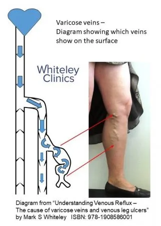
Varicose veins – diagram showing which bulging veins are visible on the surface on standing in a patient with varicose veins – The Whiteley Clinic
What causes varicose veins?
The commonest cause of varicose veins is the valves failing in leg veins. When the patient stands up, the valves let the blood fall back down the leg. As the blood should flow up the vein, this is the wrong way. Hence we call this “venous reflux”.
A valve that stops working is “incompetent”. When all the valves in a vein fail, then the vein is “incompetent”.
Therefore, when the valves fail in the superficial veins the blood can reflux into the surface veins (in the AC3). When this happens, and the veins are seen bulging on the surface. The patient has varicose veins.
Clearly, the varicose veins on the surface are not the problem. The real problem is which veins are allowing blood to reflux into them. This is why a venous duplex ultrasound scan identifies the underlying cause. It is only by treating the underlying cause that good long-term results can be obtained.
Some people ask: Can I have varicose veins I can’t see?
Yes. Venous reflux can be present in the truncal veins without any visible surface changes. In these cases, the superficial veins remain unaffected, so no bulging veins appear on the skin.
What are “hidden varicose veins”?
However, sometimes the valves only fail in one or more of the saphenous veins. As we have seen above, the saphenous veins lie in their own compartment called the AC2. These are not visible on the surface.
If the blood does not reflux into any of the veins in AC3, there will be no varicose veins to see. This happens in about 50% of people with vein disease.
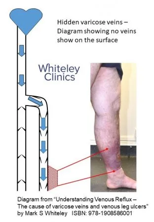
Hidden varicose veins – a diagram showing why there are no visible varicose veins on the surface on standing in a patient with venous skin damage- The Whiteley Clinic
If blood refluxes down the saphenous vein, it will hit the ankle causing inflammation. This will cause skin damage if left untreated for long enough. Initially, this is ankle oedema (swelling of the ankle – CEAP C3). This can then progress to venous eczema or brown skin changes (haemosiderin – CEAP C4). Finally, if the reflux is not treated, it is possible to develop venous leg ulcers (CEAP C6).
So what do we call reflux down the saphenous vein causing aching or one of the above problems? We cannot call it “varicose veins” as there are no bulging veins in AC3 and therefore no visible varicose veins.
In the past, this has had a variety of names. Common names are:
- superficial venous reflux (SVR)
- superficial venous incompetence (SVI)
- chronic venous incompetence (CVI)
However, these names often confuse doctors, nurses and not surprisingly patients as well.
This is why I started calling this problem “hidden varicose veins” in the early 2000s and published this name in 2011.
Many patients wonder why their legs hurt when no varicose veins are visible?
Pain can occur even in the absence of visible vein changes because raised venous pressure can affect tissues and microcirculation. This typically reflects early or non-visible venous dysfunction rather than surface vein dilation.
What Does It Feel Like to Have Hidden Varicose Veins?
The symptoms and signs of “hidden varicose veins” are the same as those for varicose veins except that no varicose veins are visible.
Hidden varicose veins cause:
- aching legs on standing
- tired legs on standing
- heavy legs on standing
- tender legs on standing
- thread veins or spider veins
- swollen ankles
- venous eczema (red stains) around ankles
- haemosiderin (brown stains) around ankles
- venous leg ulcers
- superficial thrombophlebitis (commonly called “phlebitis”)
If you have any of these symptoms or signs you need to have a venous duplex ultrasound scan. No doctor or nurse can tell you that you do not have hidden varicose veins. Only a venous duplex ultrasound scan, performed by a trained vascular technologist who can check for incompetent perforators and other rare causes of reflux, can tell you this for sure.
Understanding Venous Reflux – The Cause of Varicose Veins and Venous Leg Ulcers
History of “hidden varicose veins”.
The first reference in English language medical journals searchable on the Internet appears to be in 1970. Also, this was a leader in the British Medical Journal about “hidden perforator veins“.
That article made a sensible point about incompetent perforators. Even today, many if not most doctors do not look for or treat incompetent perforator veins. We have shown that incompetent perforator veins are associated with recurrent varicose veins. Also, we have shown that along with pelvic veins, incompetent perforator veins are a major reason why varicose veins come back in other practices.
The 1970 article pointed out the need to check for incompetent perforator veins. However, they did not have venous duplex ultrasound scanning and did not have the same understanding of hidden truncal vein reflux.
What everyone needs to know about “hidden varicose veins”.
We now know that about 15 to 20% of adults have varicose veins. However another 15 to 20% of adults have hidden varicose veins. This shows us that venous reflux disease is much more common than we thought. More importantly, everyone with any symptoms or signs of venous disease must have a venous duplex ultrasound scan performed by an appropriate specialist.
Only duplex ultrasound can reassure you that you do not have hidden varicose veins. Leaving hidden varicose veins without treatment results in a deterioration in 1 in 20 patients per year.
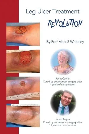
Leg Ulcer Treatment Revolution – by Prof Mark S Whiteley
Phone 0330 058 1850
Email [email protected]
Books referenced in this page:
Understanding Venous Reflux – The Cause of Varicose Veins and Venous Leg Ulcers
By: Mark S Whiteley
ISBN: 978-1908586001
Leg Ulcer Treatment Revolution
By: Mark S Whiteley
ISBN: 978-1908586056