Pigmented Purpuric Dermatosis (PPD)
Pigmented Purpuric Dermatosis (PPD) describes a group of skin conditions that may look concerning; however, they usually do not cause harm. In this article, you will discover not only what PPD is and its main symptoms, but also who most commonly develops it and the factors that trigger it.
Furthermore, we will explain when you should see a specialist, how doctors diagnose the condition, and which treatment options are available.
Finally, we will outline the possible complications if this skin condition remains untreated, ensuring you understand the full picture.

Doctors’ Definition of Pigmented Purpuric Dermatosis
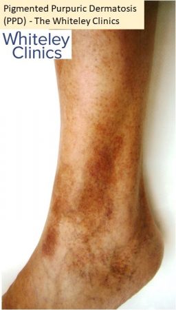
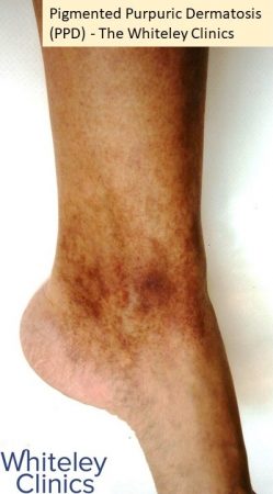
Pigmented Purpuric Dermatosis describes a group of chronic skin conditions that produce red, purple, or brown spots. These spots are usually flat and may resemble small speckles or bruises.
The colour change happens because red blood cells leak from tiny blood vessels (capillaries) beneath the skin. Subsequently, as the body breaks down these cells, iron deposits (haemosiderin) gradually create a rusty, golden-brown hue on the skin.
Although this condition may look concerning, it is generally harmless. Although it usually affects the lower legs, it may, more rarely, occur on the torso.
What Are the Symptoms and CEAP Classification?
The main symptom of Pigmented Purpuric Dermatosis (PPD) is the appearance of red, purple, or brown patches, macules, or petechiae (tiny red dots), most commonly around the ankles.
Although some patients may experience mild itching, many remain symptom-free. Moreover, even when varicose veins are not visible, you may still have “hidden varicose veins” that trigger inflammation in the skin.
Consequently, failure to identify and manage underlying varicose or hidden veins can lead to worsening venous disease. In addition, in terms of venous disease classification, PPD typically falls under the C4 category of the CEAP system (Clinical, Etiology, Anatomy, Pathophysiology), which encompasses skin changes caused by chronic venous disease, such as pigmentation and eczema.
Who Is Most Likely to Develop PPD?
Doctors consider Pigmented Purpuric Dermatosis a rare skin condition. Although it can affect people of any age, including children, it occurs more commonly in middle-aged and older adults. Moreover, men seem to develop it slightly more often than women.
Around 1 in 20 patients with C4 disease may progress to venous leg ulcers each year if underlying vein problems are not addressed.
What Causes Pigmented Purpuric Dermatosis?
In most cases, doctors cannot identify a clear cause for this skin condition, making it idiopathic. Still, a range of risk factors and causes has been identified by specialists.
- Venous problems: Varicose veins and venous insufficiency may increase pressure in the lower leg veins, leading to capillary leakage.
- Capillary fragility: Weak or fragile blood vessels can allow red blood cells to leak into the skin.
- Medications: Some medications have been linked to this skin condition, including aspirin, acetaminophen, carbamazepine, diltiazem, furosemide, interferon-alpha, and others.
- Exercise and standing: Both activities can raise venous reflux in the legs.
- Immune factors: Some cases show inflammation involving lymphocytes and macrophages, suggesting an immune-related component.
In most cases, doctors cannot identify a clear cause for this skin condition, making it idiopathic. Still, a range of risk factors and causes has been identified by specialists. Once these potential causes are understood, it is useful to recognise the main clinical types of Pigmented Purpuric Dermatosis.
Clinical Types of Pigmented Purpuric Dermatosis
There are four main subtypes of pigmented purpura, and doctors primarily recognise them based on clinical features. However, some forms also display distinctive histopathological changes:
- Progressive Pigmentary Dermatosis (Schamberg’s disease):
In some instances, the condition may progress slowly over time, a form known as Schamberg disease. Characterised by reddish-brown patches on the lower legs. - Purpura Annularis Telangiectodes of Majocchi:
Presents as ring-shaped or annular patches of purpura with visible dilated capillaries, typically seen in younger patients. - Lichen Aureus:
Appears as a solitary golden or rust-coloured plaque, most commonly located on one leg. It usually remains localised and persistent. - Pigmented Purpuric Lichenoid Dermatitis of Gougerot and Blum:
It is characterised by a combination of pigmented purpura and lichenoid papules, which give the skin a slightly raised and inflamed appearance.
When Should You See a Specialist?
Although Pigmented Purpuric Dermatosis is usually harmless, you should see a doctor if:
- The spots spread quickly or change in appearance.
- The itching becomes severe or persistent.
- You may experience varicose veins or other signs of venous reflux.
- You are concerned about the cosmetic appearance.
The best specialists to consult are dermatologists and vascular specialists; however, in some cases, your GP may initially examine you and then refer you to the appropriate expert. If you are experiencing any of the symptoms mentioned, it’s best to book a free consultation.
How Do Doctors Diagnose This Skin Condition?
In most cases, diagnosis relies on evaluating the clinical presentation of the lesions. Doctors can often recognise Pigmented Purpuric Dermatosis by its typical red-brown patches and distribution on the legs.
In certain cases, additional tests may be needed:
- Duplex Doppler ultrasound: This test checks for venous insufficiency or varicose veins in the legs, which may contribute to PPD.
- Skin biopsy: A small sample of skin may be taken to rule out other conditions, such as vasculitis or cutaneous lymphoma.
- Blood tests: Complete blood count (CBC) and clotting studies help exclude clotting disorders or low platelet counts.
The Role of Duplex Ultrasound in Diagnosis
A venous duplex ultrasound plays a crucial role when doctors assess skin changes around the ankles. Consequently, a specialist vascular technologist performs this test, as it requires expertise to identify both obvious and subtle venous problems.
Furthermore, their careful evaluation ensures that even minor issues, which a quick scan might miss, are detected and properly assessed. At The Whiteley Clinic, our technologists follow The Whiteley Protocol, examining not only the main deep and superficial veins but also the smaller perforating veins that can often be overlooked during a brief scan by a doctor.
In some patients, doctors may identify hidden varicose veins or incompetent perforating veins. When they find these, they can treat them effectively using The Whiteley Protocol. However, patients with Pigmented Purpuric Dermatosis usually have normal main leg veins.
Thanks to the high-definition capability of duplex ultrasound, specialists can sometimes visualise the tiny veins directly beneath the skin discolouration, which standard scans might miss.
Additionally, the scan may reveal a small amount of fluid in the tissue, called oedema. When oedema appears, it suggests that the veins might be leaking, which in turn provides further insight into the underlying condition.
Treatment Options for Pigmented Purpuric Dermatosis
In most cases, Pigmented Purpuric Dermatosis is harmless and does not require medical intervention. However, some patients seek treatment to relieve discomfort, such as itching, aching, or swelling, while others are concerned about the appearance of their skin.
The safest and simplest approach is wearing compression stockings. These carry minimal risk and can reduce symptoms effectively. In addition, bioflavonoid supplements combined with vitamin C may support capillary health and improve skin condition.
For patients experiencing more pronounced inflammation or irritation, topical corticosteroid creams of medium to high potency can be applied to relieve redness and itching. In selected cases, doctors may also prescribe oral medications such as griseofulvin, colchicine, or pentoxifylline to manage symptoms.
Microsclerotherapy may be recommended when cosmetic improvement is the main concern. This treatment targets underlying small veins that contribute to skin discolouration. All interventions require careful medical supervision to ensure safety and effectiveness.
At centres such as The Whiteley Clinic, specialists tailor each treatment plan to the individual. They monitor progress closely and adjust management as needed. By combining lifestyle measures, supplements, topical or oral medications, and, when appropriate, cosmetic procedures, patients can manage symptoms safely and improve the appearance of their skin.
What Can Happen If This Condition Is Misdiagnosed
Even though treatment can manage symptoms, misdiagnosis can still pose challenges. Although Pigmented Purpuric Dermatosis remains benign, it can persist for months or even years if left untreated. Moreover, the pigmentation often fades very slowly, and the condition may flare up again.
In addition, the primary concern involves appearance, as visible patches can distress some patients. Occasionally, PPD can resemble or even precede more serious conditions, such as cutaneous T-cell lymphoma. Therefore, specialists advise regular follow-up to monitor any changes and ensure early detection.
Final Thoughts
Pigmented Purpuric Dermatosis is a harmless yet often persistent skin condition that primarily affects the legs. It produces reddish-brown spots due to leaking capillaries and iron deposits within the skin. Although treatment is not always required, several options exist to relieve symptoms and improve appearance. Consulting a dermatologist or vascular specialist ensures accurate diagnosis and effective management.
With appropriate information and support, patients can manage PPD confidently and minimise its impact on daily life.