Can Microsclerotherapy Effectively Remove Spider Veins?
July 18, 2026
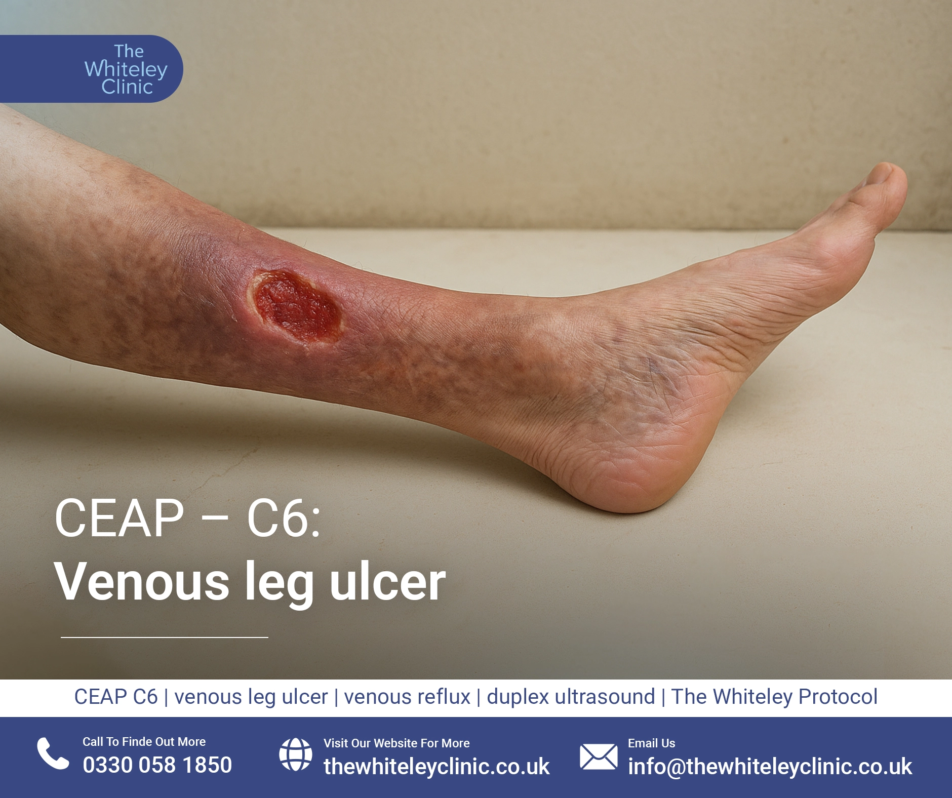
In the CEAP classification of venous disease, the worst clinical class is C6. The C6 classification means active venous leg ulcer.
An ulcer is a break in the skin that will not heal. Many non-medical people often call this a “sore” which does not seem to heal. However when there is a break in the skin which does not heal simply, the medical name for this is an ulcer. Of course, ulcers are not only restricted to legs and can occur elsewhere. For example, a stomach ulcer is when the lining of the stomach breaks down and there is a non-healing area where the stomach acid can attack the underlying muscle causing pain.
When an ulcer occurs in the leg, it is obviously called a leg ulcer. Leg ulcers are relatively common and in the UK, approximately 500,000 people get recurrent leg ulcers. At any one time approximately 125,000 people have open leg ulcers. The others are usually treated by dressings and compression alone, which result in them temporarily healing but coming back again in the future. With proper investigation and treatment by a specialist vein clinic such as The Whiteley Clinic, most leg ulcers can be permanently cured.
The important thing to know about a leg ulcer is what is the underlying cause. The major causes of leg ulceration are venous problems (usually venous reflux where the blood falls down the veins due to faulty valves, although occasionally venous obstruction due to blocked veins can be the problem), followed then by the much less common arterial problems for example, diabetes, pressure, vasculitis and malignancy.
Fortunately, for people with leg ulcers, approximately 60 to 80% of leg ulcers have an underlying vein problem. In the majority of these, the underlying vein problem can be fixed and so the leg ulcer should be curable.
It is essential to know the difference between:
The Whiteley Clinic always attempts a permanent cure for all patients with leg ulcers and our published research shows that we achieve this in approximately 85% of our patients
Venous leg ulcers can look quite different in different people and different situations. Generally they happen around the lower leg, usually just above the ankle bone although they can be at the ankle bone or even below that and onto the foot. Sometimes they are so large they go right around the leg and spread from above the ankle bone to below the ankle bone. In other cases, they can be very small and appear just in one place.
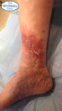
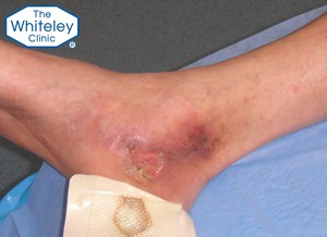
Healing venous leg ulcer after endovenous surgery under local anaesthetic at The Whiteley Clinic – CEAP-C6
Usually, venous leg ulcers are described as being on the inner side of the lower leg. However, they can occur on the outside of the lower leg or the front of the lower leg. Rarely, they can occur at the back of the lower leg.
The ulcer itself appears to be an open sore with no skin covering the underlying tissue. Depending on how clean or otherwise the ulcer is, it can appear pink and healthy, or it can have yellow green debris within it and sometimes, if it has been allowed to dry, they can be a single or multiple scabs over the top of it.
The surrounding skin is usually red as the body is using inflammation to try to heal the ulcer. In a great many cases, there is also brown skin around the ulcer. The usual progression of venous disease is red and brown skin damage around the lower leg (see CEAP C4) which if not treated, can progress to C6 leg ulceration. As such, the brown staining is usually still obvious.
In about half of the patients with a venous leg ulcer, there are visible varicose veins somewhere on the leg, usually on the calf or thigh above the ulcer. In the other half, there are no visible varicose veins but the varicose veins are hidden deep under the skin where they can only be seen with venous duplex ultrasonography.
Finally, many patients with venous leg ulcers also have dark “venous flares” which are like dark spider veins or thread veins around the inner ankle bone and stretching down onto the foot. Occasionally these can appear to be “blue blebs”.
Regardless of what is seen, anybody who has a venous leg ulcer should be referred to a vascular service, if it has been present for two weeks or more. Since July 2013, this has been official NICE recommendations in the UK.
A normal leg has skin covering it. When a leg ulcer occurs, it means that something has gone wrong that stops the normal process of skin growing and repairing on the leg.
Venous leg ulcer curable by endovenous surgery using The Whiteley Protocol® – CEAP-C6
Although many patients will put this down to a cat scratch or other trauma, of course this is not the case. In normal people, any minor trauma such as a scratch or a bump would heal. In patients where a minor scratch or trauma goes on to a leg ulcer, something is going on within the patient to stop the skin healing normally.
Hence there is always an underlying cause as to why a leg ulcer occurs.
In the majority of people who develop leg ulcers, the problem is due to veins in the legs and pelvis not working properly. Fortunately, the usual problem is that the valves inside the veins are not working, allowing the blood to fall the wrong way down the veins, hitting the ankle from the inside and causing damage. This is called “venous reflux” and the same problem can cause leg thread veins (CEAP C1), varicose veins (CEAP C2), swelling of the ankles (venous oedema CEAP C3) and skin damage of the lower legs (CEAP C4).
Fortunately, superficial venous reflux can be identified by specialise venous duplex ultrasonography and in virtually every case, can be successfully treated under local anaesthetic with the new endovenous procedures formalised under The Whiteley Protocol®.
As has been noted before, superficial venous reflux can only be seen as varicose veins in 50% of patients who suffer from it. The other 50% do not know they have it until they develop one of the problems above or a leg ulcer. In these patients, we often say they have “hidden varicose veins” although the proper medical terms vary widely and include terms such as “superficial venous reflux” (SVR), “superficial venous incompetence” (SVI) or even “chronic venous incompetence” (CVI).
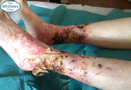
Severe venous leg ulcers drying out during elevation – CEAP-C6
If you want to understand venous reflux more and how it causes these venous problems, please see the book “Understanding venous reflux – cause of varicose veins and venous leg ulcers”.
Less commonly, the veins may be blocked. This often occurs due to a deep vein thrombosis (DVT) or can be a rarer problem such as May-Thurner syndrome. The Whiteley Clinic specialises in such conditions using up-to-date investigations such as intravascular ultrasound (IVUS)
Finally, although the majority of patients are venous in nature, arterial insufficiency (blocked or narrowed arteries) and other rarer conditions can cause leg ulcers. Of course these would not then be “venous leg ulcers” and so would not be classified under the CEAP C6 classification, as the CEAP classification is only for venous disease. However, the specialists at The Whiteley Clinic are all trained to identify leg ulcers that are not venous in origin and in many cases, using the duplex ultrasound scanner, we are able to diagnose exactly what the underlying cause is, even if it is not venous.
If you do have a venous leg ulcer, or indeed any leg ulcer and do not know the underlying cause, then you should have a venous duplex ultrasound scan if it hasn’t disappeared within two weeks. If it has disappeared within two weeks of forming, it is not an ulcer but a wound that has healed.
Once a leg ulcer has been present for two weeks or more, then a referral to a specialist vascular service such as The Whiteley Clinic is required. This has now become the clinical guidelines from the National Institute of Health and Clinical Excellence (NICE CG 168).
Before 1986, leg ulcers were treated with compression bandaging and dressings. Research from 1986 showed that the majority of venous leg ulcers were due to venous reflux (varicose veins or hidden varicose veins) and so were curable. As such, since that time, anyone who knows about varicose veins and venous research has been performing venous duplex ultrasound scans on all of their patients with venous leg ulcers, and then treating any reflux that is found. It is by following this protocol that The Whiteley Clinic has managed to cure 85% of the patients with venous leg ulcers that attend
See our published research and also the PubMed published research for more information.
Unfortunately, it has taken until July 2013 for this to become the NICE guidelines and even today, despite these guidelines, many patients with venous leg ulcers continue to be treated with compression bandaging and dressings and do not get referred for a venous duplex ultrasound scan and expert diagnosis from a vascular service such as The Whiteley Clinic. The result of this is that patients get temporary healing, and then a few weeks to months after the bandages and dressings are removed, the ulcer recurs because the underlying problem has not been fixed.
It is important to note that the recommendations from the NICE guidelines is that any “vascular service” performing such investigations and treatments should be performed by a multidisciplinary team that includes a duplex ultrasound scan and has access to all the different treatment modalities. A single doctor performing their own scan and then offering a treatment does not comply with the NICE CG 168.
Therefore to cure the ulcer permanently, the underlying vein problem needs to be identified by a venous expert such as a consultant at The Whiteley Clinic, using the results from the specialist venous duplex ultrasound scan performed by a The Whiteley Clinic technologist and then a curative treatment planned following The Whiteley Protocol®.