

How Can You Effectively Treat Calluses and Corns on Feet?
August 15, 2026
Calluses and corns on feet may begin as harmless thickened skin. However, if left untreated, they often cause pain, discomfort, and mobility issues. Consequently, addressing them early can prevent further complications.
This article explains why these lesions develop, then details how The Whiteley Clinic diagnoses and treats them. Finally, it outlines what patients can expect during follow-up and prevention.
Calluses are areas of hard, thickened skin that result from chronic mechanical stress—friction or pressure, that the foot experiences regularly. The skin responds by creating a tougher barrier.
You often see calluses over weight-bearing zones: the heel, the ball of the foot, or over bony prominences rubbing against footwear or adjacent toes.
Consequently, corns develop as calluses with an intensified centre, becoming more conical, dense, and pressing deeply into the underlying soft tissue. Several types exist, including:
When the hardened tissue presses into the soft tissues beneath, it causes pain ranging from mild discomfort to severe sensitivity. For this reason, safe and effective treatment must first address the pressure source before removing the hardened tissue.
Any effective treatment strategy for calluses and corns on feet must rest on three pillars:
Prior to removing calluses and corns, the underlying cause of pressure must be resolved. Otherwise, the hard tissue will simply recur.
Moreover, the corn or callus develops as a natural defence mechanism to protect the soft tissue beneath from ongoing irritation. If the hardened layer is removed without correcting the pressure point, the exposed healthy tissue can suffer direct damage, causing pain, inflammation, and rapid reformation of the lesion.
Therefore, effective treatment always starts with removing the cause before removing the corn or callus itself.
If the cause of pressure isn’t addressed, even expertly removed corns or calluses will often reappear.
At The Whiteley Clinic, a comprehensive, patient-centred protocol integrates diagnosis, precise intervention, and preventive strategies. The following outlines the step-by-step approach:
Initial evaluation includes foot mechanics, gait analysis, footwear history, and structural factors such as toe deformities or bone prominence.
In addition, pressure mapping or imaging sometimes complements the assessment.
This approach identifies not only the lesion but also its underlying cause.
Calluses are composed of non-living, thickened skin.
Sterile scalpel blades pare away layers in very thin slices until the lesion is resolved.
Meanwhile, they preserve a thin protective layer to prevent exposing raw tissue.
Since the outer layers lack nerve endings, the process is painless and typically does not cause bleeding.
Usually, one session suffices. Follow-up occurs after 6 to 8 weeks to monitor for recurrence or further therapy.
For hard corns, first remove the surrounding callus, then perform enucleation to extract the dense central core.
Consequently, during healing, fill the cavity with a soft gel, silicone, or polymer to offload pressure and help prevent recurrence.
In more complex cases, especially when a bony driver contributes, core removal can be combined with minimally invasive structural correction. Local anaesthesia ensures patient comfort during the procedure.
Soft corns form in moist, inter-toe spaces. After debridement, insert a toe separator to keep the toes apart, reduce friction, and promote airflow. Additionally, maintain clean and dry web spaces. To further reduce moisture, apply surgical spirit (a mild alcohol solution).
Seed corns are tiny, clustered lesions often associated with dry skin (anhidrosis). First, carefully remove the small lesions. Then, apply emollient creams, often containing urea, to maintain skin hydration and flexibility.
A review is scheduled after about 3 months to monitor for further lesions.
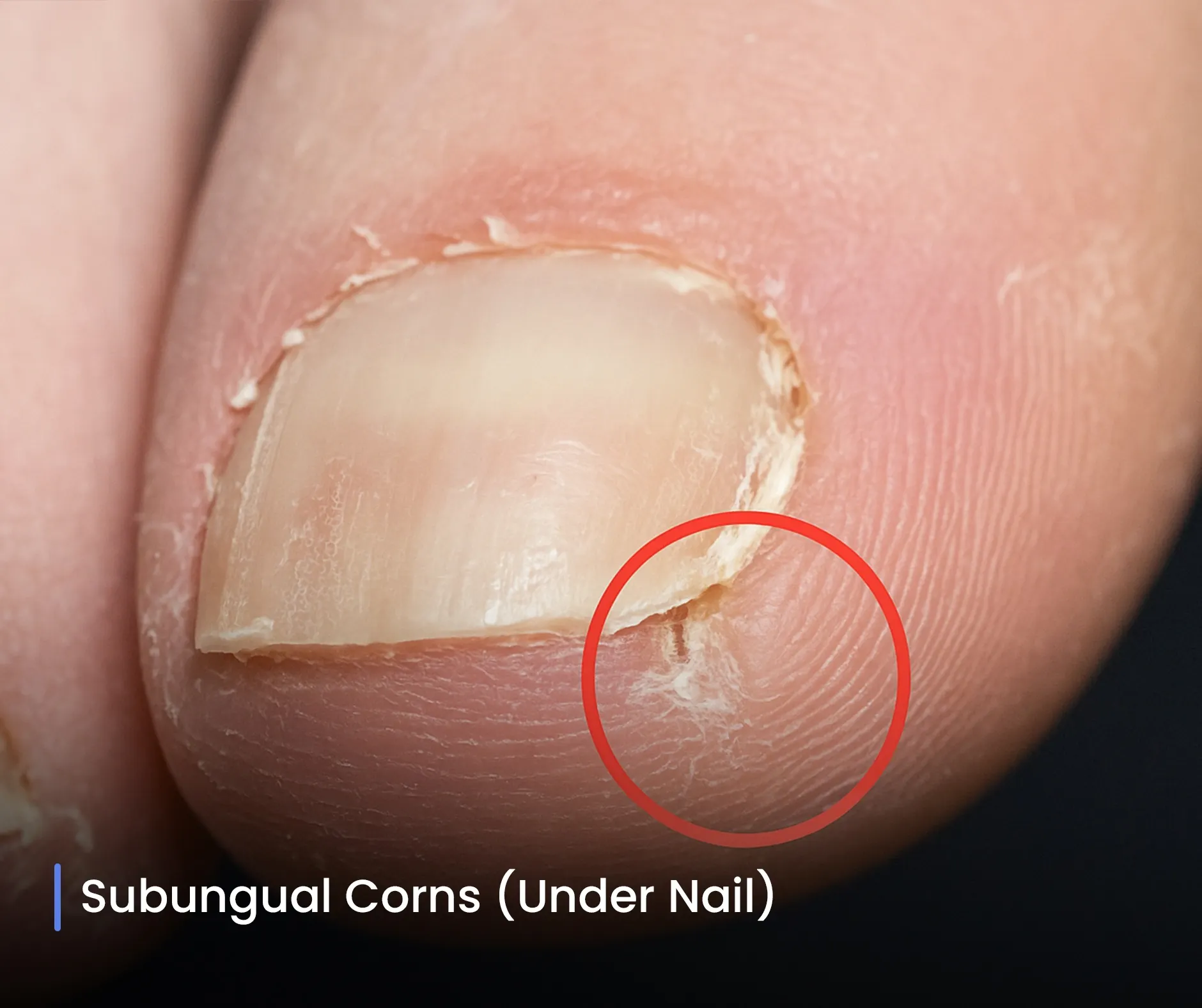
When a corn lies beneath a toenail, first trim or partially lift the nail to access the lesion safely. In some cases, temporarily remove the entire nail. After removing the corn, apply a caustic agent such as silver nitrate to destroy any residual lesion cells and reduce recurrence. Then, schedule follow-up every 2–3 weeks to monitor nail regrowth and check for new lesions.
These rare lesions contain both nervous and vascular tissue, which makes treatment more delicate. Therefore, use local anaesthesia to minimise pain and bleeding. Next, carefully excise the lesion, and then apply a caustic agent if needed to eliminate any residual tissue. Finally, schedule frequent follow-up, often weekly when caustics are used, to monitor healing and prevent recurrence.