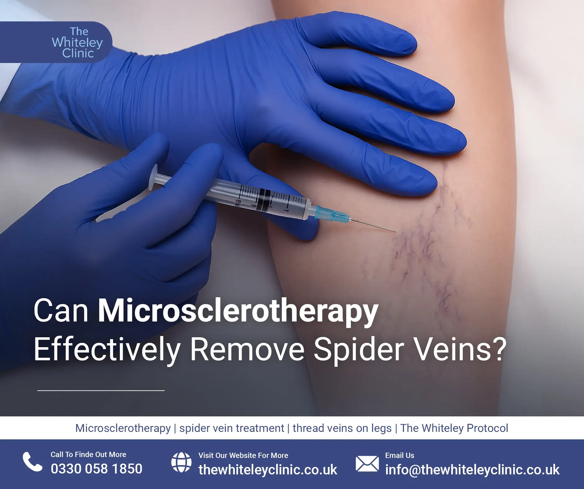
Can Microsclerotherapy Effectively Remove Spider Veins?
July 18, 2026
The classification of varicose veins into C2A or C2S is a very subjective classification and not based on much science. The main reason to separate varicose veins (CEAP C2) into asymptomatic varicose veins CEAP C2A and symptomatic varicose veins CEAP C2S, is to give a classification for private medical insurance companies, government health departments and other funders of health, to decide which patients with varicose veins should get treated and which should not.
The difficulty with symptoms is that the are felt by the patient. Some patients may say they feel symptoms when they do not, hoping to get treatment. Other patients have had the symptoms for so long, they do not realise they have the symptoms until the symptoms have been removed. This is much like wearing an uncomfortable or itchy shirt. Sometimes the worst symptoms are when you remove it!
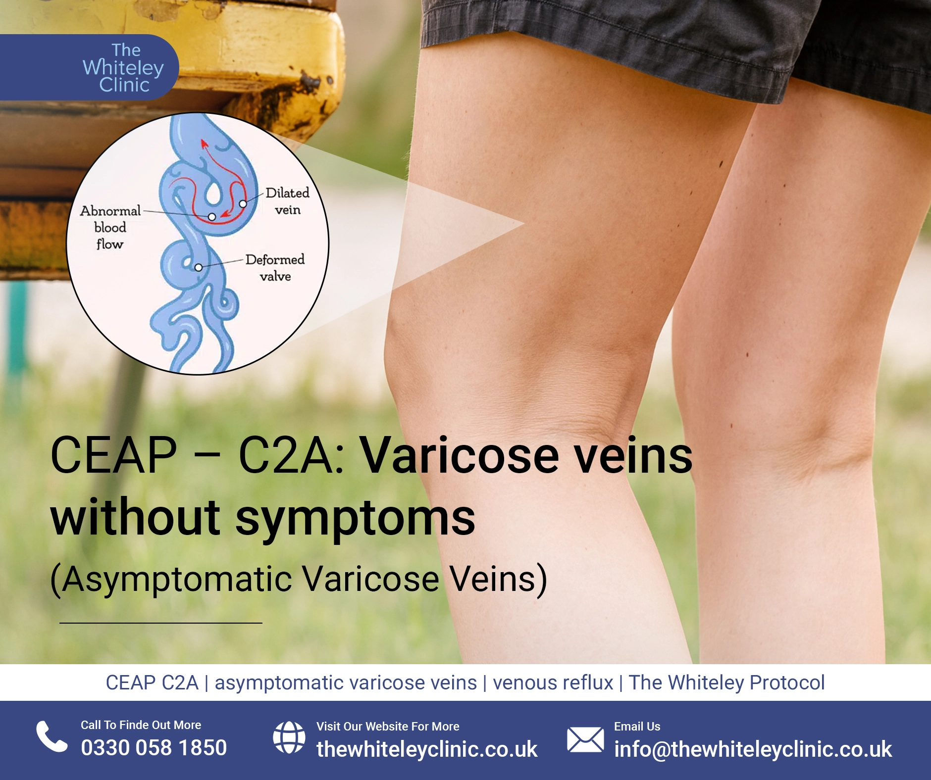
Varicose veins classified CEAP C2A are leg varicose veins that can be seen but do not cause any symptoms to the patient. Patients often say these are only cosmetic. It is important that they have not got any brown stains, red stains or have any other skin changes around them or else they become CEAP C4 (skin damage) even if the patient doesn’t feel any discomfort.
Visually, CEAP C2A varicose veins look identical to CEAP C2 varicose veins. There are bulging varicose veins on the legs on standing which can be skin coloured, blue or green. They reduce in size and can even disappear on lying down and elevating the leg.
As the distinguishing factor to make varicose veins classified as CEAP C2A, is the lack of any symptoms, there is nothing visually that can distinguish CEAP C2A as a subcategory of CEAP C2.
As CEAP C2A varicose veins are just CEAP C2 varicose veins which do not cause any symptoms to the patient, it is not surprising that the underlying causes for CEAP C2A varicose veins are exactly the same as those for CEAP C2 varicose veins.
When varicose veins first appear, they start by being simple down notations on the surface veins lower in the legs (see: Understanding Venous Incompetence book). As they become more established and larger, they are associated with underlying reflux in the majority of cases. Traditionally, this was thought to be incompetence or reflux in the great saphenous vein or small saphenous vein. However it is now known that this can also be due to incompetence in bifid sections of these veins, anterior accessory saphenous vein, Giacomini vein, incompetent perforators and pelvic vein reflux.
It has also become clear that failure to identify and treat all of the sources of venous reflux underlying a tragic varicose veins will lead to inadequate treatment and either varicose veins remaining there after treatment or, if they temporarily disappear due to post-operative bruising and thrombosis, early recurrence.
Many people decide not to do anything, particularly in the short term, if they have asymptomatic varicose veins. If the varicose veins are truly asymptomatic, there is no swelling of the ankles and no skin damage or itching, it is possible to leave the varicose veins alone at the current time although, it will continue to deteriorate. As varicose veins are actually a sign of something else going on in the venous system, the deterioration may be that the varicose veins just look worse with time, or they may go on to become symptomatic, bleed, develop clots (superficial thrombophlebitis or “phlebitis“), cause ankle swelling (CEAP C3), cause skin damage (CEAP C4) or even leg ulcers (CEAP C6). The chances of this deterioration depend on what the underlying venous problem is. As such, many people decide to get asymptomatic varicose veins treated to either prevent further deterioration or because they do not like the look of varicose veins.
Even if a person is unsure as to whether they want to have the veins treated, it is sensible to get an expert venous duplex ultrasound scan performed of the underlying veins, to find out what the risks are of further deterioration. In 2006, there was a randomised controlled research study published proved that if uncomplicated varicose veins are treated surgically, the patient had a better quality-of-life two years later than if they were only prescribed compression stockings. See: www.ncbi.nlm.nih.gov Therefore, there is good research evidence showing that patients with uncomplicated varicose veins should have them investigated and treated.
Once a venous duplex ultrasound scan has been performed by a qualified specialist vascular scientist, a full and frank discussion can then be had as to what treatment would be required if the veins were to be treated. This might be as simple as some foam sclerotherapy or phlebectomy if only the superficial veins are involved, but often underlying veins such as the great saphenous, anterior accessory saphenous or small saphenous vein have become incompetent requiring endovenous laser, radiofrequency, clarivein or glue treatment. In addition, approximately 40% of people will have incompetent perforators if a scan is performed correctly by a fully trained vascular scientist and one in seven women will have pelvic varicose veins causing the leg varicose veins.
The key to excellent treatment and long-term results is to have a specialised venous duplex ultrasound scan performed by trained specialist who looks at all of these areas and make sure none of these are missed. This is why The Whiteley Clinic only trust venous duplex ultrasound scans performed by their own specially trained specialist vascular scientist who have not only undergone formal training as vascular scientists but have then undergone training in The Whiteley Protocol® on top of their basic training and experience.
Once the true pattern of venous disease is established, treatment can be tailored to each patient and their own individual venous reflux problem.